Recent studies have debated whether alpha oscillations are crucial for how the brain processes sensory information through periods of varying excitability. To investigate, this #EEGManyLabs project is replicating a key study by Mathewson et al. (2009), which found that the visibility of visual stimuli and the amplitude of visual evoked potentials like N1, P1, P2, and P3, depend on their timing relative to alpha phases. If the replication is successful, a new experiment will explore if these effects rely on the predictability of the stimulus onset time.
M Ruzzoli, M Torralba, N Molinaro, C Benwell, D Berkowitz, D Brignani, L Falciati, L Greenwood, A Harris, C Huber-Huber, B Jack, C Keitel, M Kopčanová, C Madan, K Mathewson, S Mishra, S Mishra, P Morucci, N Myers, N Myers, F Nannetti, S Nara, J Pérez-Navarro, T Ro, N Schaworonkow, J Snyder, S Soto-Faraco, N Srinivasan, D Trübutschek, U Ajmeria, A Zazio, F Mushtaq, Y Pavlov, D Veniero "An #EEGManyLabs study to test the role of the alpha phase on visual perception (a replication and new evidence)." OSF, 2023. https://osf.io/3dhpx
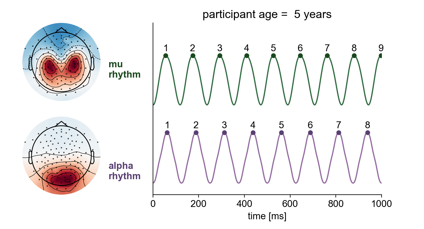 In the field of brain rhythms, one of the rock-solid empirical findings is that oscillatory frequency increases across development, for alpha and mu rhythms. In this new 〰️preprint〰️, we also look at waveform shape across development & neurodevelopmental disorders.
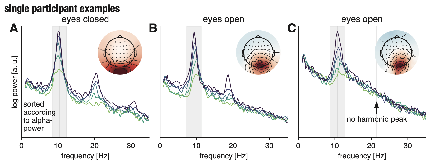 Beta-band activity in the human cortex as recorded with noninvasive electrophysiology is of diverse origin. In addition to genuine beta-rhythms, there are numerous nonsinusoidal alpha-band rhythms present in the human brain, which will result in harmonic beta-band peaks. This type of activity has different temporal and response dynamics than genuine beta-rhythms. Here, it is argued that in the analysis of higher-frequency rhythms, the relationship to lower-frequency rhythms needs to be clarified. Only in that way we can arrive at strong, methodologically valid interpretations of potential functional roles and generative mechanisms of neural oscillations.
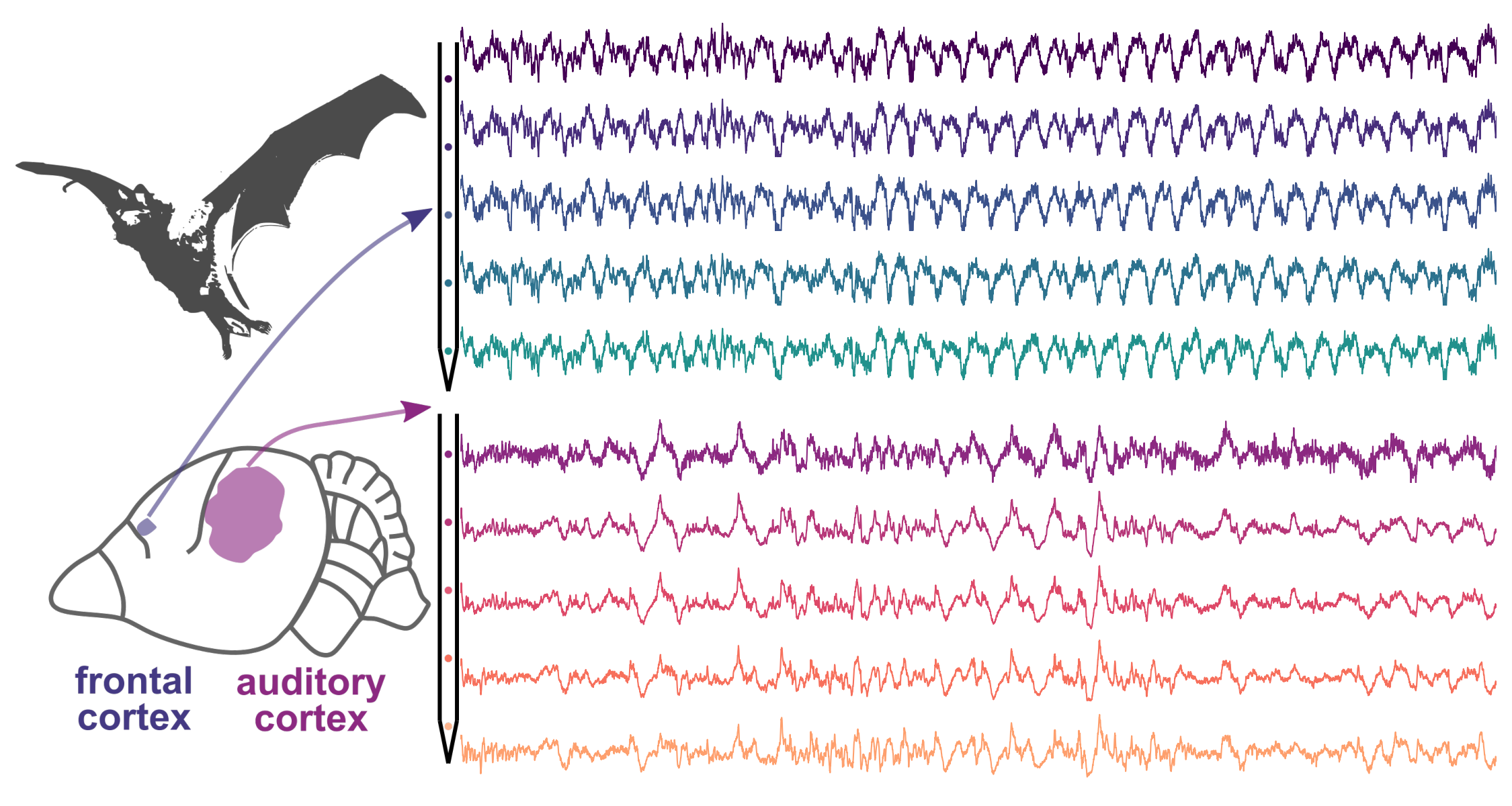 A fun collaboration with my colleague Francisco: This study investigates how the waveform shape of neural oscillations varies between different but functional related cortical areas in the brain, using simultaneous local field potential (LFP) recordings in the auditory and frontal cortices of awake bats. It finds significant differences in waveform shape, even for rhythmic activities of similar frequency, between these regions. Additionally, the study observes consistent variability in waveform shape across individual cycles and a higher correlation between spikes and LFPs in regions with more asymmetric waveforms, particularly in the frontal cortex. These findings suggest that oscillatory activity dynamics are distinct in different cortical areas, reflecting the anatomical and functional diversity of neural circuits.
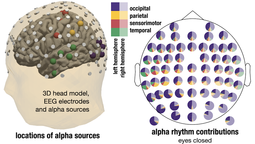 Using simulations and some EEG & MEG data, we visualized how sensor space activity results from the contribution of many different types of alpha-rhythms. Visual alpha rhythms are strong and also contribute a lot of activity of frontal sensors, the extent of it may be surprising.
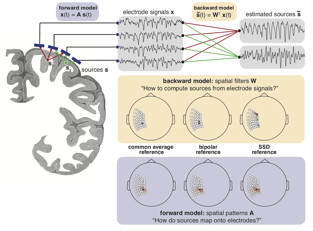 I got to look at a rare type of recordings from the brain, intracranial EEG. Because there are many different types of oscillations & other types of activity present at a given moment in time, they may overlap in space and time. Here we explore if there is a better way of extracting oscillations in this type of data, trying to reconstruct activity generators from the signal recorded on electrodes.
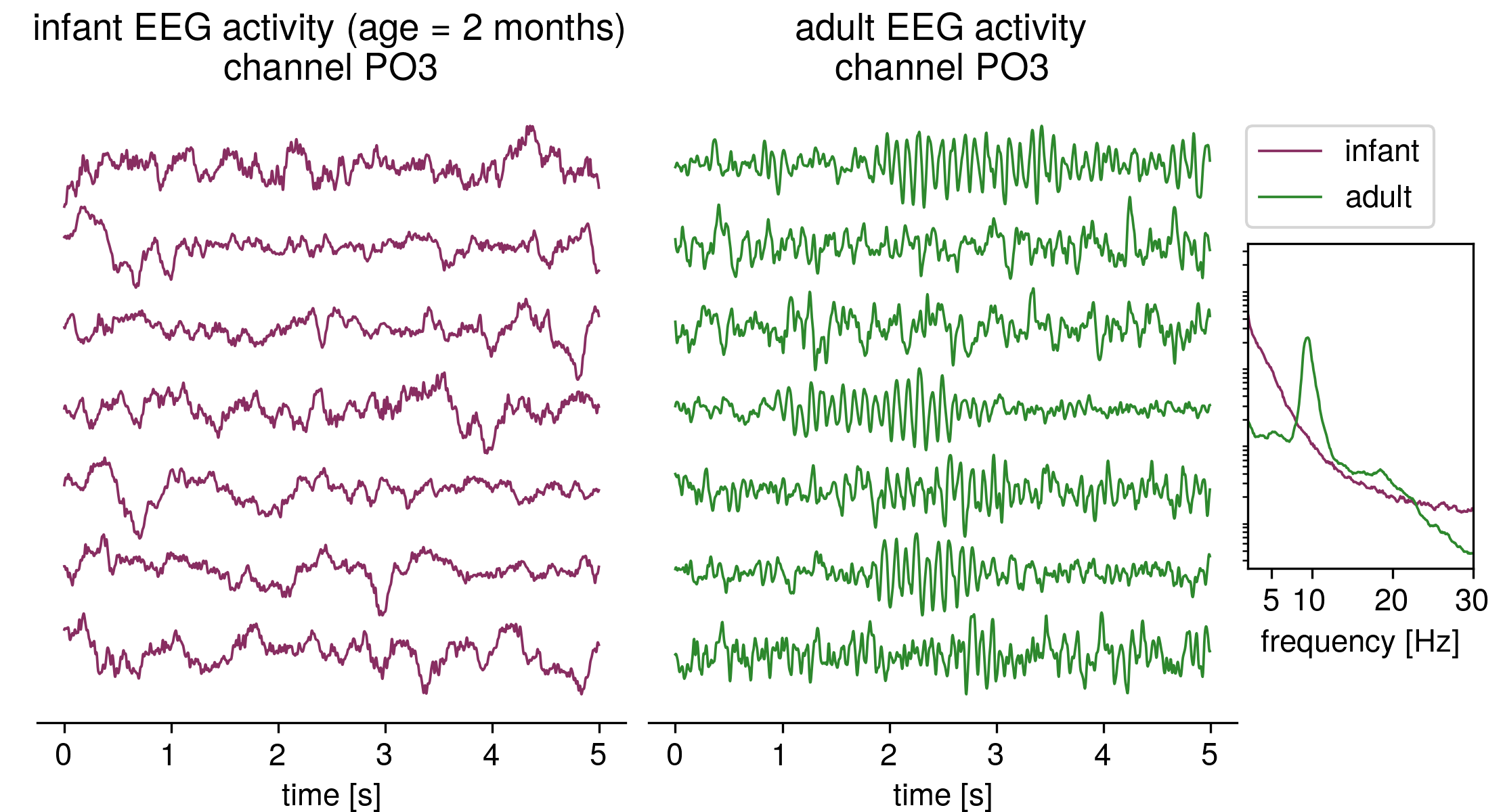 Adults have pronounced alpha-oscillations in the EEG, but young infants lack them. Oscillations emerge only gradually during the first year of life. so, in the EEG power spectrum of a 2-month year old baby, there are no pronounced peaks, they show a 1/f-power spectrum, where the spectral power decreases with increasing frequency. In this article, we looked at changes to this type of aperiodic activity across age and found a very robust decrease in the aperiodic exponent. Quanta Magazine features our work in a popular science article describing this type of brain activity:
 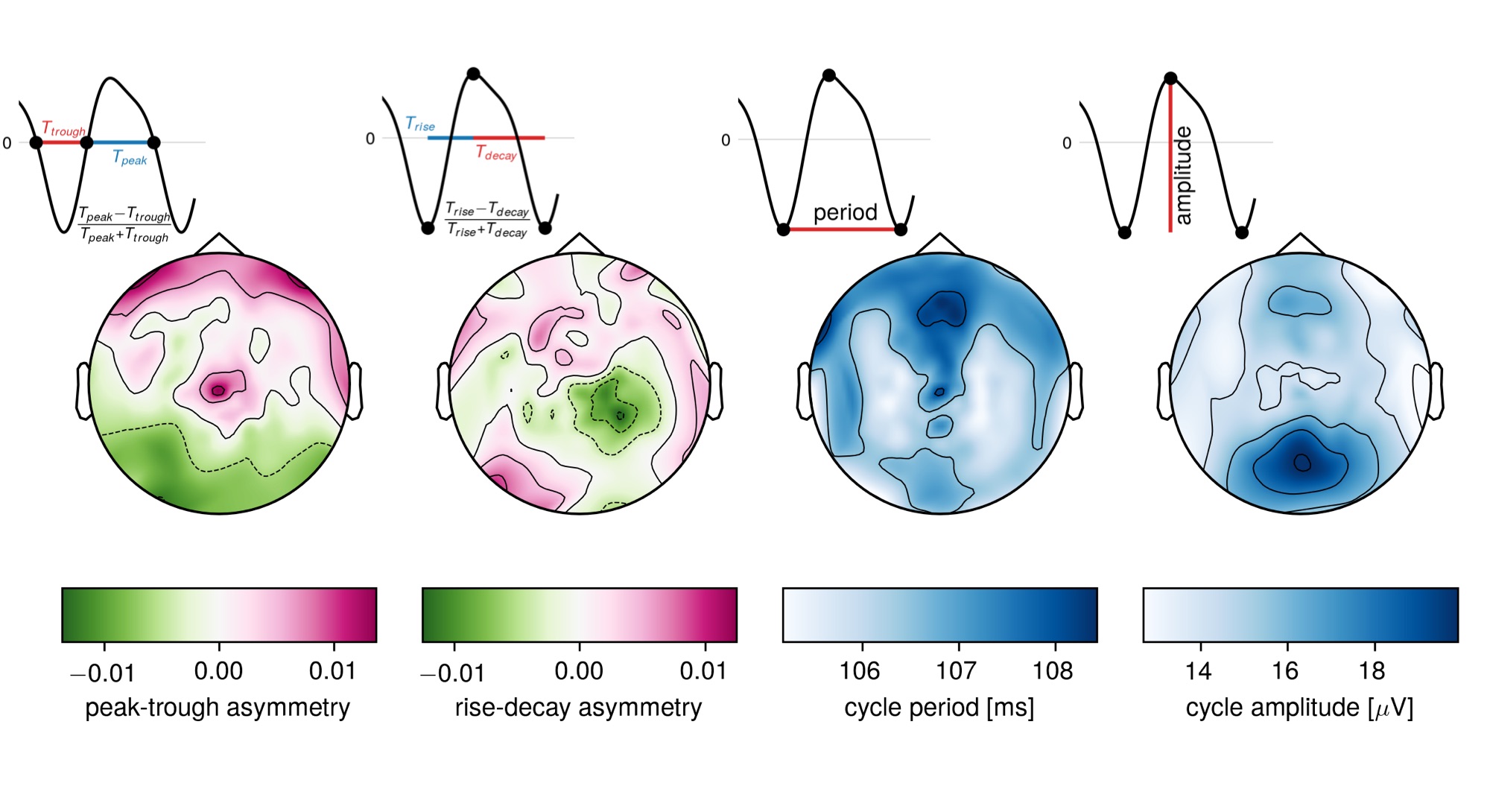 EEG/MEG oscillations have been treated as sinusoids up until recently. But they feature some beautiful waveforms, strongly deviating from a sine wave. In this article with my colleague Vadim Nikulin from the Max-Planck-Institute in Leipzig, we proposed measures of waveform shape in the time domain and explored these measures in a large EEG dataset.
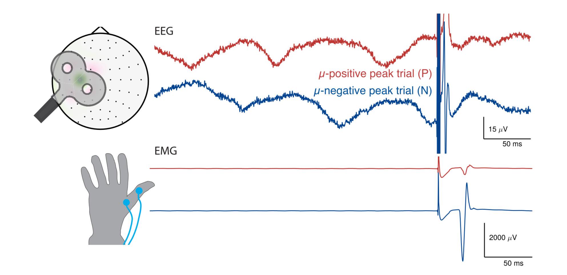 Ongoing brain activity influences behavior, but most non-invasive research in that direction relies on correlational analysis. With transcranial magnetic stimulation, a causal investigation is possible. A large portion of my PhD was concerned with brain-state dependent stimulation: we apply stimulation depending on the phase of the sensorimotor rhythm and observe large modulation of evoked muscle responses depending on the phase. Interestingly, in experiments this is only the case for some subjects, so the factors necessary for the modulation still need to be uncovered.